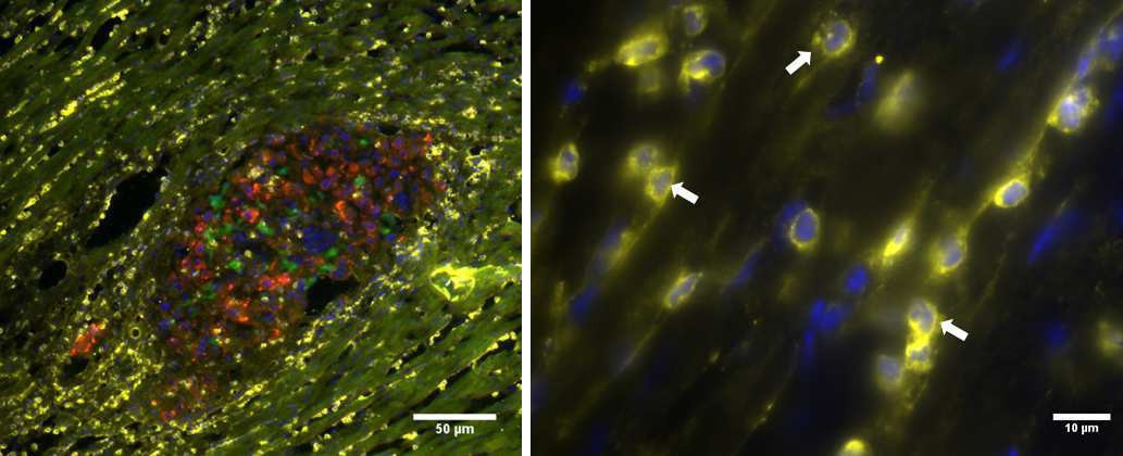
Fig. 6. Histological observation of neutrophil invasion. A tissue section from a healthy heart transplanted with ES-CMs (eGFP-expressing: green) and MSCs (red) was stained with antibodies against neutrophils (yellow). Nuclei were stained by Hoechst dye (blue). Arrows (right panel) point to multi-lobed nuclei.